
Narcolepsy type 1 may affect how fluid moves through the brain: Study
Link found between water channel protein, signaling molecule tied to sleep
Written by |

People with type 1 narcolepsy (NT1) may have changes in how their brain fluid barrier functions, a new study suggests.
Lower levels of the water channel protein aquaporin-4 (AQP4), responsible for water balance and perivascular fluid movement (the flow of cerebrospinal fluid through spaces surrounding blood vessels in the brain), were associated with having less hypocretin-1, a signaling molecule that plays a key role in sleep-wake cycles. People with NT1 have a near-complete loss of hypocretin-producing nerve cells.
“These findings suggest changes in the function of the brain-fluid-barrier in NT1, suggesting a potential … link between AQP4 as a mediator of perivascular fluid movement and hypocretinergic dysfunction,” researchers wrote.
The study, “Aquaporin-4 in Narcolepsy Type 1: Investigation of Perivascular Fluid Movement in Sleep Disorders,” was published as a brief communication in the Annals of Clinical and Translational Neurology.

NT1 patients had significantly lower median AQP4 levels
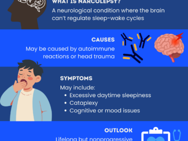
Narcolepsy affects the brain’s ability to regulate sleep-wake cycles, resulting in symptoms such as excessive daytime sleepiness and sudden sleep attacks. As a result of sleep issues, people with the disease also commonly experience cognitive issues and mood changes.
NT1 is caused by autoimmune-mediated loss of hypocretin-1 in the cerebrospinal fluid (CSF), which surrounds the brain and spinal cord. Symptoms of NT1 are generally more severe and include disrupted nighttime sleep and cataplexy, or the sudden, temporary loss of muscle tone without loss of consciousness.
In this study, a research team in Germany analyzed AQP4 levels in the CSF of people with NT1. AQP4 is notably found on astrocytes, which represent the majority of cells in the human central nervous system (brain and spinal cord) and support neurons.
The study enrolled 132 adults with NT1, whose CSF levels of hypocretin-1 were lower than 110 picograms (pg)/mL. As control groups, it also included 143 participants with other sleep disorders, matched for age and sex, and 62 without signs of neurodegenerative or neuroinflammatory disease.
Results demonstrated that NT1 patients had significantly lower median AQP4 levels than those with other sleep disorders or without sleep disorders (3.31 pg/mL vs. 4.15 pg/mL vs. 3.96 pg/mL, respectively). The difference remained significant after adjusting for patients’ age.
According to the researchers, lower AQP4 levels may reflect altered astrocyte function, lower astrocyte replacement rate (when new astrocytes replace older ones), or region-specific structural changes in these cells.
Additionally, in patients whose CSF hypocretin-1 levels were within the quantification range of a commercially available test, the amount of this signaling molecule was significantly associated with the amount of AQP4 in their CSF.
“The positive correlation of CSF AQP4 and hypocretin-1 levels with lower levels of both biomarkers in NT1 supports a possible connection of hypocretinergic neurosignalling and perivascular fluid exchange,” the researchers wrote. “These data suggest potential brain fluid barrier alterations in NT1.”



Leave a comment
Fill in the required fields to post. Your email address will not be published.