
Brain hormone may drive sudden loss of muscle tone in narcolepsy
Mouse study findings may offer new opportunities for treating cataplexy
Written by |

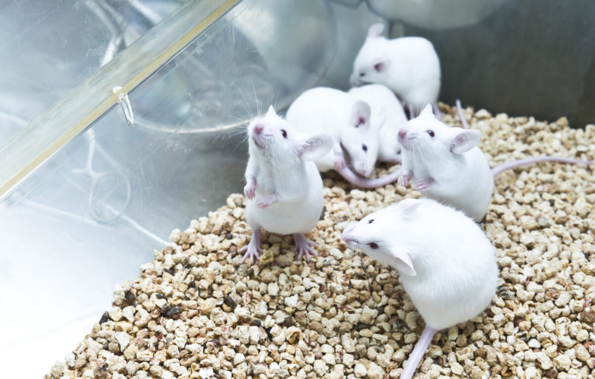
Six white lab mice huddle together in a cage.
Positive, strong emotions may trigger cataplexy, or a sudden loss of muscle tone, in people with narcolepsy by activating a brain pathway dependent on the oxytocin hormone that switches off nerve cells responsible for maintaining muscle tone, a study in mice suggests.
Researchers found that reuniting narcoleptic mice with their littermates increased cataplexy, while blocking oxytocin — a hormone involved in social bonding and reward — prevented that increase.
Experiments in the mouse model showed that oxytocin activates a specific group of nerve cells in the central amygdala, a brain region involved in processing emotions, which in turn suppress nerve cells responsible for preventing muscle tone loss. This, ultimately, allows cataplexy to occur.
Chocolate, a rewarding stimulus also associated with strong, positive emotions, activated the same oxytocin-dependent pathway.
“These findings show that oxytocin acts through the amygdala to promote cataplexy triggered by social and other rewarding stimuli, offering new opportunities for treating cataplexy,” researchers wrote.
The study, “Oxytocin promotes socially triggered cataplexy,” was published in Nature Neuroscience by a team of researchers in the U.S.

‘Social dimension of cataplexy has received little attention’
Narcolepsy is a disorder marked by symptoms such as excessive daytime sleepiness and falling asleep uncontrollably. In type 1 narcolepsy, the disease is caused by the near-complete loss of nerve cells, or neurons, that produce hypocretin, a brain signaling molecule also known as orexin that regulates wakefulness.
This type is also characterized by cataplexy, a sudden loss of muscle tone usually lasting a few minutes. Episodes are often triggered by strong positive emotions and commonly occur during social interactions, such as laughing with family or friends, playing games, or unexpectedly seeing someone close.
“Yet, this social dimension of cataplexy has received little attention,” the researchers wrote.
Previous research had already identified specific neurons in the central amygdala as a critical relay between emotional signals and brainstem circuits that control muscle tone.
However, the central amygdala contains many different types of neurons, and “the neuronal mechanisms through which social stimuli promote cataplexy are poorly understood,” the team wrote.
Because positive social interactions trigger the release of oxytocin, and oxytocin is known to activate neurons in the central amygdala, the researchers suspected that a specific population of oxytocin-responsive neurons in this brain region might be responsible for triggering cataplexy.

Reunions with other mice caused increase in cataplexy episodes
To test their hypothesis, the team turned to a mouse model of narcolepsy that lacks the gene encoding orexin. They found that animals housed together experienced about twice as many cataplexy episodes as those housed alone.
The researchers then temporarily separated littermates for 20 minutes before reuniting them. Reunion caused a sharp increase in cataplexy episodes, while animals that were not reunited with their littermates showed no change in the number of cataplexy events.
Similarly, orexin-deficient mice given chocolate in addition to their regular food experienced significantly more cataplexy than when fed regular chow alone.
The team next asked whether oxytocin was responsible. Giving orexin-deficient mice carbetocin, a drug that activates oxytocin receptors — proteins on the surface of nerve cells that bind oxytocin — increased cataplexy in a dose-dependent manner. Conversely, administration of an oxytocin receptor blocker reduced the increase in cataplexy triggered by carbetocin and social reunion.
“Oxytocin signaling is necessary and sufficient for promoting cataplexy,” the researchers wrote.
Brain oxytocin levels increased during social interactions
Further experiments showed that brain oxytocin levels increased during social interactions and rose shortly before cataplexy began. In addition, oxytocin activated a specific population of neurons in the central amygdala that have oxytocin receptors.
These oxytocin-responsive neurons became more active during social interactions and consistently increased their activity in the seconds before a cataplexy episode. The same neurons also became active before chocolate-triggered cataplexy.
Artificially activating these neurons roughly doubled the number of cataplexy episodes. Conversely, silencing them prevented the increase in cataplexy normally triggered by either social reunion or chocolate.
These findings highlight the importance of [oxytocin-responsive neurons in the central amygdala] in generating cataplexy and provide new opportunities to develop medications targeting these pathways.
Additional experiments showed that oxytocin-responsive amygdala nerve cells send suppressive signals to the ventrolateral periaqueductal gray (vlPAG), a brain region that helps prevent muscle atonia — the muscle tone loss that occurs during cataplexy. By suppressing the vlPAG, these neurons remove an important brake on muscle atonia, allowing cataplexy to occur.
Blocking the connection between the oxytocin-responsive neurons in the central amygdala and the vlPAG significantly reduced the surge in cataplexy episodes triggered by chocolate or carbetocin. This suggests that disrupting this pathway may reduce cataplexy triggered by rewarding stimuli, the researchers noted.
“These findings highlight the importance of [oxytocin-responsive neurons in the central amygdala] in generating cataplexy and provide new opportunities to develop medications targeting these pathways,” the team wrote. “Testing whether an oxytocin [blocker] reduces cataplexy in people with narcolepsy type 1 could help establish whether a similar mechanism is relevant in humans.”
But because blocking oxytocin may affect normal social behavior and have limited clinical utility, the researchers emphasized the importance of finding other ways to block these oxytocin-responsive neurons.




Rachel Nesmith
Fascinating! Loving more cataplexy scientific discoveries!