
In-ear EEG device may be alternative to laboratory sleep studies for narcolepsy
New custom-fit device found to accurately track stages of deep sleep
Written by |
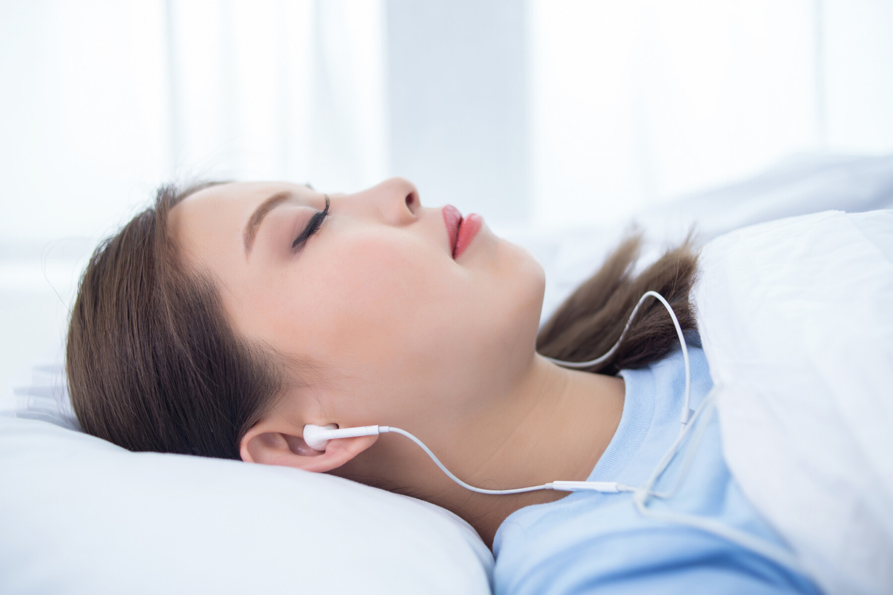
Maintaining a strict sleep schedule and a consistent daily routine can help adults with narcolepsy reduce daytime drowsiness and improve overall symptom control. (Photo by iStock)
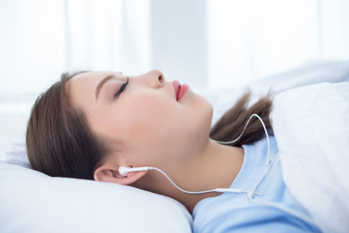
A custom-fit, in-ear electroencephalography (EEG) device that records the brain’s electrical activity may be a possible alternative to traditional in-laboratory sleep studies for diagnosing narcolepsy, a new study suggests.
According to researchers, the new, wearable EEG device showed substantial agreement with standard sleep studies for overnight sleep staging, as well as good agreement for measuring daytime sleepiness in healthy adults. The device, however, was less reliable at detecting brief, subtle sleep transitions in adults with central disorders of hypersomnolence — conditions, including narcolepsy, marked by an extreme, uncontrollable urge to sleep during the day.
“These findings support the feasibility of in-ear EEG for sleep staging and daytime sleepiness assessment in laboratory settings,” the authors wrote. The results also “motivate larger confirmatory studies — including home-based longitudinal monitoring — to establish clinical utility, particularly in populations with altered sleep architecture.” Sleep architecture refers to the structural pattern of sleep, which consists of distinct, cyclical stages that repeat throughout the night.
The study, “A novel, wearable, in-ear EEG technology to assess sleep and daytime sleepiness,” was published in the journal Bioelectronic Medicine. Four of the study’s 11 authors are employed by Nextsense, the California-based company that makes the device.

Narcolepsy is a chronic neurological condition that affects the brain’s ability to control sleep-wake cycles. It’s characterized by dysregulated nighttime sleep and overwhelming daytime sleepiness.
Sleep studies typically done with scalp sensors in specialized labs
The diagnostic workup for narcolepsy typically involves polysomnography, a sleep study that records brain activity, heart rate, breathing, and muscle movements during overnight sleep. Conducted in a specialized facility, polysomnography monitors a person’s sleep stages and helps rule out other conditions that cause sleepiness.
In-ear EEG is being studied as a possible alternative to traditional in-laboratory sleep studies, offering potential advantages of greater comfort and practicality.
Here, a research team tested the Nextsense device against polysomnography to assess its ability to classify sleep stages and measure daytime sleepiness.
The participants were 16 healthy adults without sleep disorders and eight patients with central disorders of hypersomnolence (CDH), a group of conditions that cause excessive daytime sleepiness. This included narcolepsy type 1 and type 2, as well as idiopathic hypersomnolence, when these problems are due to an unknown cause.
The Nextsense system consisted of custom-fit earbuds with electrodes inside the ear canals, 3D-printed from detailed scans of each participant’s ear to ensure comfort and stable contact. The earbuds were connected to a recording device and worn alongside standard polysomnography equipment, so both systems recorded simultaneously.
Healthy controls completed a sleep study followed the next day by four maintenance of wakefulness test (MWT) trials. The MWT measures the ability to stay awake while sitting quietly in a darkened room.
CDH patients also completed a sleep study and four MWT trials, with two in a crossover design: one day on their usual medication and one day during a medication holiday, with medications withheld for at least five days beforehand.
Three certified sleep technologists scored each of the five sleep stages: wake, N1 and N2 (light non-REM sleep), N3 (deep non-REM sleep), or REM sleep. The team also measured sleep onset latency (SOL), the time elapsed before sleep onset. REM, or Rapid Eye Movement, sleep is characterized by darting eye movements and increased brain activity.

In-ear EEG device showed mostly good agreement with lab tests
Among the healthy controls, the results showed substantial agreement between in-ear EEG and scalp-placed EEG, as done in traditional sleep studies. Agreement was highest for wake (90%), then N2 (84%), REM (71%), N3 (64%), and N1 (54%). N1, the lightest and most transitional sleep stage, was the hardest to score reliably with either method.
For overall sleep architecture, the two devices showed strong agreement for total sleep time, sleep efficiency — the proportion of time actually spent asleep — and wake after sleep onset, which is time spent awake after first falling asleep.
Across all 126 MWT trials with patients and controls, overall agreement in SOL between the two devices was moderate. In 37 of the trials in which both devices detected sleep, agreement was good.
In healthy controls, when both detected sleep, agreement was excellent. In the eight CDH patients who completed two MWT days each, agreement between the in-ear and scalp EEG devices for SOL was moderate overall. The in-ear device recorded a mean SOL that was 10.1 minutes longer than the scalp EEG, a statistically significant difference, the researchers noted.
In 22 trials, scalp EEG detected sleep, but the in-ear device did not. The weaker agreement between in-ear and scalp EEG in CDH was driven mainly by 15 trials from five of the CDH patients, in which scalp EEG alone detected sleep. This involved predominantly brief, subtle transitions into N1 sleep.
Overall, the in-ear EEG device detected sleep in CDH trials less often than in healthy control trials (55% vs. 73%). When SOL was averaged across all four trials per participant, the standard clinical approach, agreement was good across all 24 participants, “suggesting that the device captures clinically meaningful between-subject differences in daytime sleepiness despite trial-level discordances,” the team wrote.
In the CDH crossover study, 7 of 8 patients showed a longer in-ear SOL on their medication day compared with their medication-holiday day, with a mean increase of 8.83 minutes. The two patients with narcolepsy type 1 showed the smallest improvements, specifically 1.84 and 4.68 minutes.
“The present findings reflect a controlled, in-laboratory evaluation of in-ear EEG against concurrent gold-standard [polysomnography],” the team wrote, noting that “these results compare favorably to prior ear-EEG sleep-staging validation work.”
Still, “generalization to broader clinical populations and to unsupervised home-based deployment will require larger confirmatory studies,” the researchers wrote.



Leave a comment
Fill in the required fields to post. Your email address will not be published.